Orthodontic treatments are constantly evolving, thanks in part to technological advancements that make treatment more comfortable and gentle for patients. The intraoral scanner is a perfect example, as it often makes taking impressions, which are generally felt to be unpleasant, obsolete.
However, i’m frequently asked why an orthodontist continues to use radiography in the context of modern orthodontic treatment, especially in a city like Geneva. In patients’ minds, radiography is often seen as harmful or “outdated.”
Yet, a complete diagnosis is essential for the success of orthodontic treatment. Without proper radiographic examination, such diagnosis is impossible, as the orthodontist needs to evaluate the state of the teeth and jaw.
What is a cephalometric x-ray?
Orthodontic treatment always requires a complete diagnosis. It’s the only way to precisely plan the exact course, necessary measures, and approximate timeline of the orthodontic treatment.

At Dental Geneva, for children and teenagers, i always perform a cephalometric x-ray of the skull that provides a complete image of the skull from the front and side and offers an overview of the patient’s dentition and jaws. This allows me to “predict” the type of skull growth (either in length or width) and its possible consequences on teeth occlusion.
It also enables me to assess the position of the lower and upper jaws as well as the position of the front teeth, which influences the final treatment plan.
This type of image can therefore be taken from childhood. It allows for a reliable prognosis for the future development of the child’s jaw and to establish whether or not there is a need for a removable or fixed dental appliance.

What is a panoramic x-ray?
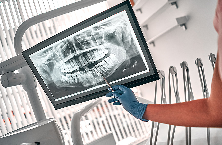
A panoramic x-ray, also known as an orthopantomogram (OPT), on the other hand, provides the ability to determine the current state of the jaw and skull just before a possible orthodontic treatment.
With an OPT radio, i can particularly observe
- The placement of teeth in the jaw
- The condition of the dentition
- Tooth malformations
- Inflammations of the tooth root and the jawbone
The images are renewed during treatment, to monitor the position of the dental axes and to record changes in the appliance and the tooth root.
These two types of radiographs are the foundation of a successful orthodontic treatment. They are an additional control instrument for the orthodontist. Radiographs not only allow for precise monitoring of the treatment’s progress but also enable timely corrections as necessary.

Digital radiography: the gentle method!
At dental Geneva, we use only digital radiography. Its radiation exposure is roughly at the level of natural background radiation and is generally about 90% lower than that of traditional radiographs.
This is particularly due to the brief time needed to take the images. The actual shooting lasts only a few seconds, and the transfer to the computer, hence digital radiography, takes about a minute. Despite the brevity of the shooting, maximum quality is guaranteed. The images are even of better quality and more detailed than with the traditional method.
Digital shooting also allows for trouble-free transmission of images and avoids the costly evaluation and development of images in a dark room.
We can view the images with you without delay on the treatment room screens. For further processing, the contrast of the images can also be easily adjusted.
Thus, digital radiography is faster, gentler, and more efficient than the traditional analog method.
Our team also takes many protective measures to reduce the impact of radiation, however slight it may be.
Still have questions about digital radiography?
If you have any questions about digital radiography, do not hesitate to contact us! At our dental Geneva clinic, we are always at your disposal to inform you about this topic or others. Thanks to modern equipment, we guarantee orthodontic treatment that is as gentle and considerate as possible.


